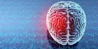
An engineering team used piezoelectricity to create wearable sensors that evaluate eye movement in order to diagnose brain illnesses or brain injury. A team of engineers at the University of Houston has created wearable sensors that can study eye movement to diagnose brain illnesses or injury. Many brain disorders and abnormalities manifest as eye symptoms, frequently before other symptoms occur.
You see, the eyes are more than just a doorway into the soul, as poets claim. These extremely valuable organs serve as an extension of the brain, providing early warning symptoms of brain-related illnesses as well as information on their causes. Examination of the eyes can also aid in tracking the progression and symptoms of physical and mental shocks to the brain.
Researchers say current eye-tracking systems have flaws and deliver insufficient amounts of data. Plus, they’re bulky, with multiple electrodes on the face and neck, expensive and have weak outputs.
We believe that the F-PEMSA can be employed in many clinical studies concerning brain disorder conditions such as ADHD, autism, Alzheimer’s disease and Parkinson’s disease as well as the aftermath of traumatic brain injuries like post-concussion syndrome and post-traumatic stress disorder.
Jae-Hyun Ryou
And in the blink of an eye…improvement
The new method, developed in the UH lab of Jae-Hyun Ryou, associate professor of mechanical engineering, with assistance from Nam-In Kim, postdoctoral researcher, is non-invasive, comfortable to wear, and safe, allowing for easy and continuous measurements and monitoring of eyeball movements when combined with a hand-held display and computing device.
The new sensors are elegant and flexible, comprised of a thin, crystal-like sheet that creates electricity when bent or moved. The piezoelectricity effect is a phenomenon that causes certain materials to create an electric charge in response to mechanical stress. The output voltages from top, middle, and lower sensors, or transducers, on distinct temple locations produce discernible electrical patterns.
“Skin-attachable wearable sensors for monitoring vital signs and biomedical parameters are components of great importance in personal healthcare and portable diagnostic systems,” reports Ryou in Advanced Healthcare Materials. “Among them, thin-film piezoelectric sensors offer unique advantages of easy fabrication at low cost, a wide range of available sizes, lightweight, excellent mechanical flexibility and stability, rapid reaction rate, high sensitivity, high signal-to-noise ratio and excellent long-term stability and durability.”
“The new sensors are easy to wear and can be used in brain-eye relationship studies to evaluate the brain’s functional integrity,” he said.
Intense focus on disease
Ophthalmological examinations of eye blinking patterns have been used to diagnose stroke, multiple sclerosis, Parkinson’s disease, and Alzheimer’s disease in the early stages. Ocular motions are also significantly linked to a variety of brain illnesses, as brain function influences eyeball and upper eyelid control.
Previous research examined abnormal blink rate and blink modulation in children with attention-deficit hyperactivity disorder, with the spontaneous blink serving as a measure of the dopaminergic system’s integrity in the brain. Autism has also been linked to motor neurons in the brain that control the movement of the eyes and muscles.
“We believe that the F-PEMSA can be employed in many clinical studies concerning brain disorder conditions such as ADHD, autism, Alzheimer’s disease and Parkinson’s disease as well as the aftermath of traumatic brain injuries like post-concussion syndrome and post-traumatic stress disorder, potentially offering the prospect of early and accurate diagnoses and the development of personalized therapies,” said Ryou.