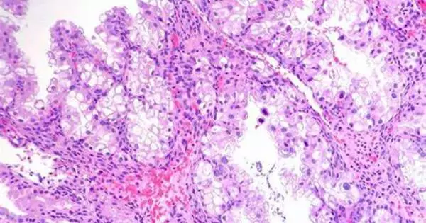
The Arias-Stella reaction is a histopathological change that occurs in the endometrium (uterine lining) during pregnancy. Arias-Stella phenomenon is a benign endometrial change associated with the presence of chorionic tissue. This reaction is named after two pathologists, Javier Arias-Stella and César Arias-Stella, who described it for the first time in 1954.
The Arias-Stella reaction is primarily caused by progesterone. It appears to be a malignancy cytologically, and it was previously diagnosed as endometrial cancer. The presence of chorionic (related to the chorion, a membrane that surrounds the fetus and contributes to the formation of the placenta) tissue within the endometrium is typically associated with this reaction.
Here are the key features of the Arias-Stella reaction:
- Appearance of Cells: The endometrium’s glandular epithelial cells undergo transformation as part of the reaction. These cells’ appearance changes dramatically.
- Nuclear Changes: The presence of enlarged, bizarre-looking nuclei in glandular cells is the most distinguishing feature of the Arias-Stella reaction. Because of their unusual appearance, these nuclei may be misdiagnosed as cancerous changes.
- Eosinophilic Cytoplasm: The cytoplasm of affected cells becomes eosinophilic, staining more intensely with eosin, a type of dye used in histology.
- Association with Pregnancy: The Arias-Stella reaction is most commonly observed in the endometrium of pregnant women, especially in cases where there is the implantation of chorionic tissue within the endometrial glands. It is often seen during the first trimester of pregnancy.
- Mimicking Malignancy: The nuclear and cellular changes seen in the Arias-Stella reaction can be mistaken for malignancy, and it is not uncommon for pathologists to encounter difficulty in distinguishing between the reaction and cancerous changes.
Significance
It is significant only because it has the potential to be misdiagnosed as cancer. It can occur in a perfectly normal pregnancy. Despite its ability to mimic malignancy, recognizing the distinguishing features and understanding the clinical context (pregnancy) can aid in distinguishing it from cancerous changes.
Diagnosis
It has nuclear enlargement and may also have an irregular nuclear membrane, granular chromatin, centronuclear vacuolization, and pseudonuclear inclusions.