About Cornea
Definition
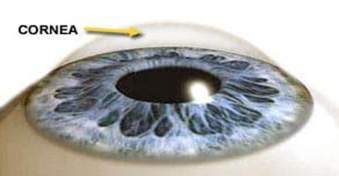
Cornea is the clear, domeshaped surface that covers the front of the eye and it is the eye’s outermost layer. It lies directly in front of the iris and pupil, and it allows light to enter the eye. The cornea is more than a protective film; it is a fairly complex structure that has five layers. The cornea’s main function is to refract, or bend, light. The cornea is responsible for focusing most of the light that enters the eye.
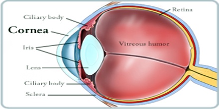
In humans, the refractive power of the cornea is approximately 43 dioptres. While the cornea contributes most of the eye’s focusing power, its focus is fixed. The curvature of the lens, on the other hand, can be adjusted to “tune” the focus depending upon the object’s distance. Medical terms related to the cornea often start with the prefix “kerat-” from the Greek word κέρας, horn.
Structure and Functions of Cornea
Cornea is composed of proteins and cells. It does not contain blood vessels, unlike most of the tissues in the human body. Blood vessels may cloud the cornea, which may prevent it from refracting light properly and may adversely affect vision. The horizontal diameter of the cornea typically measures about 12 millimeters (mm), and the vertical diameter is 11 mm, when viewed from the front. But if viewed from behind, the cornea appears circular, with a uniform diameter of approximately 11.7 mm. This makes the cornea about two-thirds the size of a dime. The center thickness of the average cornea is about 550 microns, or slightly more than half a millimeter.
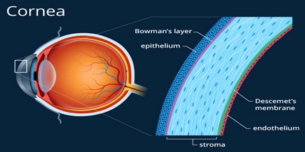
The cornea has five layers. These five layers are:
- The Corneal Epithelium: The epithelium is the cornea’s outermost region, comprising about 10 percent of the tissue’s thickness. Epithelial cells are constantly being produced and sloughed off in the tear layer of the surface of the eye. The turnover time for the entire corneal epithelium is about one week.
- Bowman’s Layer: This is a very thin (8 to 14 microns) and dense fibrous sheet of connective tissue that forms the transition between the corneal epithelium and the underlying stroma.
- The Corneal Stroma: This middle layer of the cornea is approximately 500 microns thick, or about 90 percent of the thickness of the overall cornea. It consists primarily of water (78 percent) and collagen (16 percent), and does not contain any blood vessels. Collagen gives the cornea its strength, elasticity, and form. The collagen’s unique shape, arrangement, and spacing are essential in producing the cornea’s transparency.
- Descemet’s Membrane: Under the stroma is Descemet’s membrane, a thin but strong sheet of tissue that serves as a protective barrier against infection and injuries. Descemet’s membrane is composed of collagen fibers and is made by the endothelial cells that lie below it. Descemet’s membrane is regenerated readily after injury.
- The Corneal Endothelium: This is the innermost layer of the cornea. The back of the endothelium is bathed in the clear aqueous humor that fills the space between the cornea and the iris and pupil. The corneal endothelium is only a single layer of cells thick and measures about 5 microns. Most of the endothelial cells are hexagonal (six-sided), but some may have five or seven sides. The regular arrangement of these cells is sometimes called the endothelial mosaic.
The cornea tends to repair itself quickly from minor abrasions. However, deeper abrasions may cause scars to form on the cornea, which causes the cornea to lose its transparency, leading to visual impairment. The clear cornea allows light to enter the eye for vision. But it has another very important function as well the cornea provides approximately 65 to 75 percent of the focusing power of the eye.
Reference: masseyeandear.org, healthline.com, allaboutvision.com, wikipedia.