Wearable sensors that use ultrasound technology to provide cardiac imaging are available. These sensors can provide real-time cardiac imaging without the need for traditional imaging methods such as X-rays or CT scans. This technology can be particularly useful for people who require continuous monitoring of their heart function, as it provides a non-invasive and portable solution. However, it’s important to note that the accuracy and reliability of these wearable sensors may vary, and it’s always best to consult a medical professional for an accurate diagnosis.
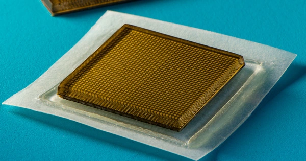
Engineers and doctors have created a wearable ultrasound device that can evaluate both the structure and function of the human heart. The portable device, about the size of a postage stamp, can be worn for up to 24 hours and is effective even during strenuous exercise.
The goal, according to Sheng Xu, a professor of nanoengineering at the University of California San Diego who is leading the project, is to make ultrasound more accessible to a larger population. Echocardiograms, or ultrasound examinations of the heart, currently necessitate highly trained technicians and large devices.
“The technology enables anybody to use ultrasound imaging on the go,” Xu said.
Thanks to custom AI algorithms, the device is capable of measuring how much blood the heart is pumping. This is important because the heart not pumping enough blood is at the root of most cardiovascular diseases. And issues with heart function often manifest only when the body is in motion. The work is described in the issue of the journal Nature.
The rising risk of heart disease necessitates more advanced and comprehensive monitoring procedures. By providing more detailed information to patients and doctors, continuous and real-time cardiac image monitoring has the potential to fundamentally optimize and reshape the paradigm of cardiac diagnoses.
Sheng Xu
Cardiac imaging is a critical clinical tool for assessing long-term heart health, detecting problems as they occur, and caring for critically ill patients. This new non-invasive wearable heart monitor for humans provides real-time, automated insights into the heart’s difficult-to-capture pumping activity, even when a person is exercising.
The wearable heart monitoring system employs ultrasound to continuously capture images of the four chambers of the heart from various angles, and then analyzes a clinically relevant subset of the images in real-time with custom-built AI technology. The project builds on the team’s previous advances in deep tissue wearable imaging technologies.
“The rising risk of heart disease necessitates more advanced and comprehensive monitoring procedures,” Xu said. “By providing more detailed information to patients and doctors, continuous and real-time cardiac image monitoring has the potential to fundamentally optimize and reshape the paradigm of cardiac diagnoses.”
Existing non-invasive methods, on the other hand, have limited sampling capabilities and provide limited data. Xu’s team’s wearable technology enables safe, non-invasive, and high-quality cardiac imaging, yielding images with high spatial resolution, temporal resolution, and contrast. “It also reduces patient discomfort and overcomes some limitations of noninvasive technologies like CT and PET, which may expose patients to radiation,” explained Hao Huang, a Ph.D. student in the Xu group at UC San Diego.
The unique design of the sensor makes it ideal for bodies in motion. “The device can be attached to the chest with minimal constraint to the subjects’ movement, even providing a continuous recording of cardiac activities before, during, and after exercise,” said Xiaoxiang Gao, a postdoctoral researcher in the Xu group at UC San Diego.
The importance of cardiac imaging
Cardiac diseases are the leading cause of death in the elderly and are becoming more common in the young as a result of lifestyle factors. The symptoms of cardiac diseases are transient and unpredictable, making them difficult to detect. This has increased demand for more advanced, inclusive, non-invasive, and cost-effective monitoring technologies, such as long-term cardiac imaging, which is made possible by this wearable device.
How it works in detail
The new system collects data via a wearable patch that is as soft as human skin and is designed for maximum adherence. The patch is about the size of a postage stamp, measuring 1.9 cm (L) x 2.2 cm (W) x 0.09 cm (T). It sends and receives ultrasound waves, which are used to generate a continuous stream of images of the heart’s structure in real-time. This ultrasound patch is soft and stretchy, and it adheres to human skin well, even during exercise.
Using ultrasound, the system can examine the left ventricle of the heart in separate bi-plane views, producing more clinically useful images than was previously available. As a use case, the team demonstrated imaging of the heart during exercise, which is not possible with the rigid, cumbersome equipment used in clinical settings.
The heart’s performance is defined by three factors: stroke volume (the volume of blood pumped out by the heart each beat), ejection fraction (the percentage of blood pumped out of the left ventricle of the heart each beat), and cardiac output (the volume of blood the heart pumps out every minute).
Xu’s group created an algorithm to enable continuous, AI-assisted automatic processing. “A deep learning model automatically segments the shape of the left ventricle from continuous image recording, extracting its volume frame by frame and yielding waveforms to measure stroke volume, cardiac output, and ejection fraction,” Mohan Li, a master’s student in the Xu group at UC San Diego, explained.
“Specifically, the AI component consists of a deep learning model for image segmentation, an algorithm for calculating heart volume, and a data imputation algorithm,” explained Ruixiang Qi, a master’s student in the Xu group at UC San Diego. “Based on the shape and area of the left ventricle segmentation, we use this machine learning model to calculate heart volume. The deep learning model for imaging segmentation is the first to be used in wearable ultrasound devices. It enables the device to provide accurate and continuous waveforms of key cardiac indices in various physical states, including static and after exercise, something that has never been done before.”
Thus, this technology can generate curves of these three indices continuously and noninvasively, as the AI component processes the continuous stream of images to generate numbers and curves.
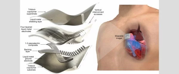
The team faced some technical challenges while developing the platform, which necessitated careful decision-making. The researchers used a piezoelectric 1-3 composite bonded with Ag-epoxy backing as the material for transducers in the ultrasound imager to produce the wearable device itself, reducing risk and improving efficiency over previous methods. They achieved superior results by selecting a wide-beam compounding transmission configuration for the transducer array. They also chose FCN-32, which achieved the highest possible accuracy, from nine popular models for machine-learning-based image segmentation.
In the current iteration, the patch is connected through cables to a computer, which can download the data automatically while the patch is still on. The team has developed a wireless circuit for the patch, which will be covered in a forthcoming publication.