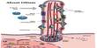
Microscopy is a scientific technique that involves using microscopes to study and analyze objects that are too small to see with the naked eye. It is the technical field of utilizing microscopes to observe items and regions of objects that cannot be seen with the naked eye (objects that are not beyond the resolution range of the normal eye).
There are three well-known areas of microscopy: optical, electron, and scanning probe microscopy, as well as the developing subject of X-ray microscopy. Microscopes use lenses or combinations of lenses to magnify and resolve the features of specimens, allowing scientists and researchers to examine the structure, composition, and behavior of materials at the microscopic level.
There are several types of microscopes, each designed for specific purposes. Some common types include:
- Light Microscopes (Optical Microscopes): These microscopes use visible light to illuminate and magnify specimens. They are commonly used in biology, medicine, and materials science.
- Electron Microscopes: These microscopes use beams of electrons instead of light to achieve much higher magnification and resolution.
- Scanning Probe Microscopes (SPM): These microscopes use a physical probe to scan the surface of a specimen, measuring various properties such as height, electrical conductivity, and magnetic field.
Optical and electron microscopy both require the diffraction, reflection, or refraction of electromagnetic radiation/electron beams interacting with a specimen, as well as the collecting of dispersed radiation or another signal to generate a picture. This method can be carried out by exposing the sample to a large field of light (as in normal light microscopy and transmission electron microscopy) or by scanning a tiny beam across the sample (as in confocal laser scanning microscopy and scanning electron microscopy).
Scanning probe microscopy involves interacting with the surface of an object of interest using a scanning probe. Microscopy revolutionized biology, gave rise to the subject of histology, and is now an important method in the biological and physical sciences. X-ray microscopy is three-dimensional and non-destructive, allowing for repeated imaging of the same sample for in situ or 4D research and allowing you to “see inside” the sample before sacrificing it to higher resolution techniques. A 3D X-ray microscope employs computed tomography (microCT), which involves rotating the sample 360 degrees and reconstructing the pictures.
Microscopy is essential in a wide range of scientific disciplines, including biology, medicine, chemistry, physics, and materials science. It has made major contributions to our understanding of biological organism structure and function, material qualities, and matter behavior at the nanoscale. Microscopy method advancements continue to play an important role in scientific research and technological growth.