Researchers have created a cell-friendly method of high-fidelity bioprinting. The ink hardened into distinct geometries, including the shape of a human nose, after multiple injections of cell-based ink and a printing support. Printed cells were viable for at least two weeks. This work is a critical step toward producing lab-grown tissues and organs and, eventually, advancing regenerative medicine and animal-free medication safety testing.
What if organ damage could be healed simply by generating a new organ in the lab? Improving researchers’ ability to print living cells on demand into geometrically well-defined, soft complex 3D architectures is critical for such work, as well as for animal-free toxicological testing.
In a study recently published in ACS Biomaterials Science and Engineering, researchers from Osaka University have overcome prior limitations that have hindered cell growth and the geometrical fidelity of bioprinted architectures. This work might help bring 3D-printed cell constructs closer to mimicking biological tissue and organs.
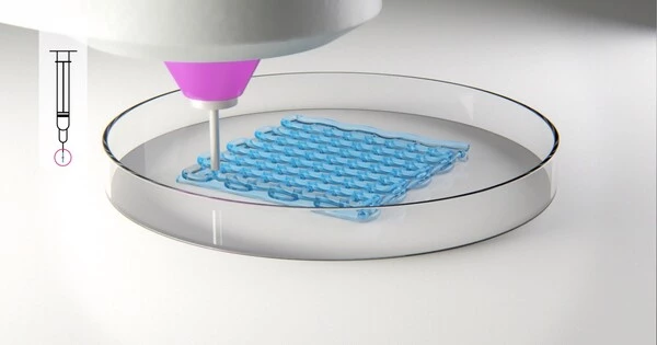
In our approach, a 3D printer alternately dispenses cell-containing ink and printing support. It’s worth noting that the support also aids in the solidification of the ink. All that is required for ink solidification is in the support, and after removing the support, the geometry of the soft-printed cell structures remains intact.
Takashi Kotani
Researchers have been exploring the possibility of this layer-by-layer tissue construction technique to rebuild damaged body parts and test medical theories since it was initially published in 1988 using a conventional inkjet printer. To build 3D structures, a cell-containing “ink” is ejected from a printing nozzle in bioprinting. Hard structures are typically easier to print than soft structures. Soft structures, on the other hand, are preferable for cell proliferation in printed structures. Printing soft structures in a printing support is effective; nevertheless, the solidification of ink in the support filled in a vessel can result in contamination of the vessel with undesired substances from the support. Ink solidification into a soft matrix using a printing support without contamination, while retaining cell viability, was the goal of this work.
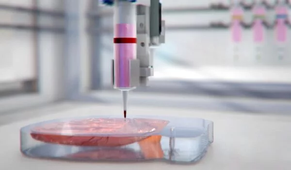
“In our approach, a 3D printer alternately dispenses cell-containing ink and a printing support,” explains Takashi Kotani, the study’s principal author. “It’s worth noting that the support also aids in the solidification of the ink. All that is required for ink solidification is in the support, and after removing the support, the geometry of the soft printed cell structures remains intact.”
The hydrogen peroxide from the support enabled an enzyme in the ink to commence gelation of the ink, resulting in a gel-enclosed cell assembly in a matter of seconds. The quick gelation prevented contamination of the assembly during creation. After removing the support, straightforward 3D constructs such as inverted trapezium geometries as well as human nose shapes– including bridges, holes, and overhangs – were readily obtained.
“We largely retain mouse fibroblast cell geometry and growth, and the cells remain viable for at least two weeks,” senior investigator Shinji Sakai adds. “These cells also adhere to and proliferate on our constructs, which highlights our work’s potential in tissue engineering.”
This novel approach represents a significant advancement in the engineering of human cell assemblies and tissues. Further research could include improving the ink and support, as well as adding blood vessels into the artificial tissue to better its likeness to physiological architectures. This work, as well as future advancements in the precision fidelity of bioprinting, will aid regenerative medicine, pharmaceutical toxicity, and other sectors.