Cherenkov imaging makes it possible to see radiation beams on a patient’s body in real time and offers a way to assess how accurately radiotherapy is administered. The quality of Cherenkov images can now be enhanced using a flexible, non-toxic sheet of carbon quantum dots (cQDs) affixed to the patient, according to Chinese researchers.
When charged particles move more quickly than the phase velocity of light in tissue, Cherenkov light is created. The exact radiation dose that is administered during therapy may be determined since the signal strength is inversely proportional to the dose provided. Comparing the optical imaging methodology to traditional techniques of radiation dose monitoring, the latter offers high spatial resolution, high sensitivity, and quick imaging speed.
However, because the Cherenkov emission is so weak, tissue scatters and absorbs the photons that are released. Standard charge-coupled device (CCD) cameras struggle to pick up the signal as a result. It is now commonplace to utilize more expensive intensified CMOS/CCD cameras.
The Cherenkov emission and absorption spectra of the cQDs overlap, and they exhibit luminescence at longer wavelengths as a result. Thus, the Cherenkov emission can be shifted to fit the ideal wavelength of a CCD camera’s sensitive detection region using the cQD sheeting, developed and proven at the Department of Nuclear Science and Technology at Nanjing University of Aeronautics and Astronautics.
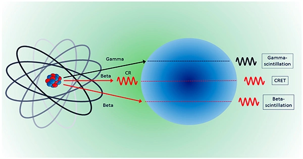
With the cQD sheeting in place, the optical emission is made up of radioluminescence produced in the cQDs, fluorescence triggered by the radioluminescence, and Cherenkov photons produced in the tissue’s superficial surface. By doing this, the total optical signal is increased, and the acquired images’ image quality and signal-to-noise ratio (SNR) are both improved.
The cQD sheeting was made by principal investigator Changran Geng and colleagues using a solution of 10 nm-diameter cQDs and UV-curable adhesive. This slurry was spun-coated onto a surface covered in plastic sheeting, and a UV lamp was used to solidify it. The plastic substrate makes sure the scintillation material doesn’t come into contact with the skin directly.
The finished cQD sheeting was flexible enough to contour to the surface of the patient and had dimensions of 15 cm in diameter and 222 5 m in thickness. The researchers observe that the cQD sheeting is nearly transparent and does not prevent tissues from emitting Cherenkov radiation.
The researchers first tested the cQD sheeting on a solid water slab covered with a 2 mm layer of light colored skin-toned clay to imitate the optical qualities of skin. They then reported their findings in Medical Physics. They used administered doses of 100–500 MU, cQD concentrations of 0, 0.05, and 0.1 mg/ml, and 6 and 10 MV beams to assess the connection between optical intensity and delivered dose. For both 6 and 10 MV photons, they noticed a linear relationship between optical intensity and dose. The SNR in both cases more than doubled with the addition of the cQD sheeting.
The team then tested the effectiveness of the cQD sheeting using various radiation materials and ambient light sources on an anthropomorphic phantom. With cQD sheeting, light emission from the surfaces of the various materials was increased by more than 60%. In particular, when cQD sheeting was added to bolus, mask sample, and a mixture of bolus and mask, the average optical intensity rose by about 69.25%, 63.72%, and 61.78%, respectively. The associated SNRs increased by roughly 62.78%, 56.77%, and 68.80%.
Optical pictures with an SNR of better than 5 could be captured through the sheeting under ambient light from a red LED. A band-pass filter was added, which raised the SNR by 98.85% roughly.
The researchers state that “the light intensity and SNR of optical pictures can be greatly boosted with a combination of cQD sheeting and matching filter.” With a quicker and less expensive picture capture procedure, this “sheds new light on the promotion of the clinical application of optical imaging to see the beam in radiotherapy.”
According to Geng, the group is aggressively pursuing its study in a variety of methods. Cherenkov imaging research for use with electron beam radiotherapy of keloids, benign fibrous lesions resulting from an aberrant healing response, is one instance.
According to some research, post-operative electron beam irradiation may lower the incidence of keloid recurrence, says Geng. “However, erroneous deliveries are frequently linked to patient setup errors, breathing movements, and variations in electron beam characteristics. These may result in an inadequate or excessive dose at the improperly spaced neighboring fields, which may harm healthy skin tissue or trigger the recurrence of keloid lesions.