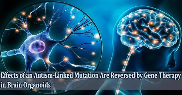
Researchers from the University of California San Diego School of Medicine used human brain organoids in a study that was published in Nature Communications on May 2, 2022, to show how a genetic mutation linked to a severe form of autism impairs neuronal development. The function of the gene was successfully restored using gene therapy methods to restore brain structure and function.
Several neurological and neuropsychiatric diseases, including autism spectrum disorders (ASD) and schizophrenia have been linked to mutations in Transcription Factor 4 (TCF4), an essential gene in brain development.
Because transcription factors control whether genes are activated or inactive, their presence or absence in the developing embryo can have a cascading effect. Still, little is known about what happens to the human brain when TCF4 is mutated.
Researchers concentrated on Pitt-Hopkins Syndrome, an ASD particularly brought on by TCF4 mutations, to investigate this issue. The majority of children with the genetic disorder are nonverbal and have severe cognitive and physical impairments.
Instead of developing a mouse Pitt-Hopkins Syndrome research model, the UC San Diego team decided to develop a human Pitt-Hopkins Syndrome research model that more closely mimics the neurological features of patients. Using stem cell technology, they converted patients’ skin cells into stem cells, which were then developed into three-dimensional brain organoids, or “mini-brains.”
Initial observations of the brain organoids revealed a slew of structural and functional differences between the TCF4-mutated samples and their controls.
What is truly outstanding about this work is that these researchers are going beyond the lab and working hard to make these findings translatable to the clinic. This is so much more than a stellar academic paper; it’s a true measure of what well-practiced science can accomplish to hopefully change human lives for the better.
Audrey Davidow
“Even without a microscope, you could tell which brain organoid had the mutation,” said senior study author Alysson R. Muotri, PhD, professor at UC San Diego School of Medicine, director of the UC San Diego Stem Cell Program and member of the Sanford Consortium for Regenerative Medicine.
Organoids with the TCF4 mutation were significantly smaller than healthy organoids, and many of the cells were actually neural progenitors rather than neurons. These unspecialized cells were originally intended to proliferate and subsequently develop into more complex brain cells, but in the mutant organoids, some aspect of this process had gone wrong.
A series of experiments revealed that the TCF4 mutation led to downstream dysregulation of SOX genes and the Wnt pathway, two important molecular signals that guide embryonic cells to multiply, mature into neurons and migrate to the correct location in the brain.
There were fewer cortical neurons created as a result of this dysregulation, which prevented neural progenitors from multiplying effectively. The cells that did develop into neurons were less excitable than usual and frequently remained grouped together as opposed to forming carefully calibrated neuronal circuits.
The flow of neuronal activity in the mutant brain organoid was disturbed by this unusual cellular architecture, which, according to the investigators, will eventually lead to compromised cognitive and motor performance.
“We were surprised to see such major developmental issues at all these different scales, and it left us wondering what we could do to address them,” said first author Fabio Papes, PhD, associate professor at the University of Campinas and visiting scholar at UC San Diego School of Medicine, who jointly supervised the work with Muotri. Papes has a relative with Pitt-Hopkins Syndrome, which motivated him to study TCF4.
For restoring the functional gene in the brain tissue, the scientists investigated two distinct gene therapy techniques. Both techniques successfully raised TCF4 levels, correcting the molecular, cellular, and electrophysiological symptoms of Pitt-Hopkins Syndrome.
“The fact that we can correct this one gene and the entire neural system reestablishes itself, even at a functional level, is amazing,” said Muotri.
Muotri points out that while in a clinical setting children would receive their diagnosis and treatment a few years later, these genetic manipulations took place at a prenatal stage of brain development.
Therefore, in order to determine whether a later intervention is still secure and efficient, clinical trials are required. In preparation for such a trial, the researchers is currently fine-tuning their recently authorized gene therapy tools, in which spinal injections of the genetic vector will presumably restore TCF4 activity in the brain.
“For these children and their loved ones, any improvements in motor-cognitive function and quality of life would be worth the try,” Muotri said.
“What is truly outstanding about this work is that these researchers are going beyond the lab and working hard to make these findings translatable to the clinic,” said Audrey Davidow, president of the Pitt Hopkins Research Foundation. “This is so much more than a stellar academic paper; it’s a true measure of what well-practiced science can accomplish to hopefully change human lives for the better.”
Co-authors include: Janaina S. de Souza, Ryan A. Szeto, Erin LaMontagne, Simoni H. Avansini, Sandra M. Sanchez-Sanchez, Wei Wu, Hang Yao and Gabriel Haddad at UC San Diego; Antonio P. Camargo, Vinicius M. A. Carvalho, Jose R. Teixeira, Thiago S. Nakahara, Carolina N. Santo, Barbara M. P. Araujo and Paulo E. N. F. Velho at the University of Campinas.
Disclosures: Alysson R. Muotri is the co-founder of and has an equity interest in TISMOO, a company dedicated to genetic analysis and human brain organogenesis.