3D printing technology is rapidly being employed in medicine, including the creation of patient-specific tumor models. A multidisciplinary multinational team of researchers has developed a way for better 3D modeling of complicated tumors.
The researchers from the University of Waterloo used cutting-edge bioprinting techniques in conjunction with synthetic structures or microfluidic chips. The technology will aid lab researchers in better understanding heterogeneous tumors, which contain more than one type of cancer cell and are frequently spread in unforeseen patterns.
Traditionally, doctors would biopsy a patient’s tumor, harvest cells, and then grow them in lab petri dishes. “For fifty years, this was how biologists understood tumors,” said Nafiseh Moghimi, a post-doctoral researcher in applied mathematics and the study’s lead author. “But a decade ago, repeated treatment failures in human trials made scientists realize that a 2D model does not capture the real tumour structure inside the body.”
Breast cancer is especially difficult to treat because it metastasizes as complicated tumors including various types of cells. Using cells from one or two biopsies to represent an entire tumor can result in inadequate treatment strategies and poor outcomes.
The team’s research addresses this issue by developing a 3D model that not only portrays the complexity of a tumor but also simulates its surroundings.
The study, conducted in the Mathematical Medicine Lab under the guidance of applied mathematics professor Mohammad Kohandel, brought together advances from numerous disciplines. “We’re doing something completely new in Canada. Perhaps only a couple of labs are doing anything remotely similar to this research,” Moghimi speculated.
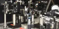
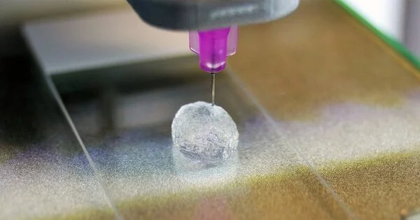
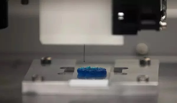
First, the researchers developed polymer “microfluidic chips”: microscopic structures etched with channels that imitate blood flow and other fluids surrounding a patient’s tumor. The researchers then cultured different types of cancer cells and suspended them in their own bioink: a cocktail of gelatine, alginate, and other nutrients designed to keep the cell cultures alive. Finally, they used an extrusion bioprinter – a device that resembles a 3D printer but for organic material – to layer the different types of cancer cells onto the prepared microfluidic chips.
Oncologists can plan and strategize treatment options using 3D-printed tumor models. These models provide a precise and realistic picture of the tumor’s location and characteristics, which is particularly useful in complex cases. These models can be used by medical professionals for training and education, allowing them to practice and perfect their abilities in a controlled environment.
As a result, scientists now have a living, three-dimensional model of complex malignancies that they can use to evaluate different ways of treatment, such as various chemotherapy medicines. Moghimi and her colleagues are particularly interested in developing sophisticated breast cancer models. Breast cancer is the second most prevalent cancer diagnosed in women, after skin cancer.
Breast cancer is especially difficult to treat because it metastasizes as complicated tumors including various types of cells. Using cells from one or two biopsies to represent an entire tumor can result in inadequate treatment strategies and poor outcomes.
The 3D-printed tumor models demonstrate how new technology allows for speedier, less expensive, and less painful therapies for major diseases such as late-stage breast cancer.